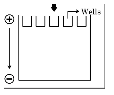
Observe the given picture carefully. A mixture of DNA with fragments ranging from 100 base pairs to 1800 base pairs were separated by electrophoresis on agarose gel with the following arrangement:
[Diagram 1 provided.]
(a) What result will be obtained in staining with ethidium bromide? Explain with reasons. [1]
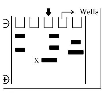
(b) The above setup was modified as shown below and a band with 100 base pairs was obtained at X.
[Diagram 2 provided.]
What changes were made to the previous design to get a band at 'X'. Why did the band appear at 'X'? [2]
Observe the given picture carefully. A mixture of DNA with fragments ranging from 100 base pairs to 1800 base pairs were separated by electrophoresis on agarose gel with the following arrangement:
[Diagram 1 provided.]
(a) What result will be obtained in staining with ethidium bromide? Explain with reasons. [1]
(b) The above setup was modified as shown below and a band with 100 base pairs was obtained at X.
[Diagram 2 provided.]
What changes were made to the previous design to get a band at 'X'. Why did the band appear at 'X'? [2]
Official answer
The separation depends on DNA being negatively charged and therefore migrating towards the positive electrode (anode).
(a) Result in Diagram 1 (on staining with ethidium bromide):
- No separated bands will be seen within the gel.
- Reason: DNA carries a negative charge (phosphate backbone) and moves towards the positive electrode. In this setup the positive electrode (+) is placed on the same side as the wells (top), so the DNA moves out of the wells/gel rather than migrating down through it — hence no proper band pattern forms even after ethidium bromide staining under UV.
(b) Changes made and why the band appears at X:
- Change made: the electrodes were reversed — the positive electrode was shifted to the bottom (and negative to the top), i.e. away from the wells.
- Why the band appears at X: now the negatively charged DNA migrates downward through the gel towards the positive electrode. Since smaller fragments move faster and farther, the smallest 100 base-pair fragment travels the farthest and appears at the lowest position, X.
(Concept note: ethidium bromide intercalates between DNA bases and glows bright orange under UV, revealing the bands once migration occurs.)
Marking scheme
- 1.(a) 1 mark: no bands seen because DNA (negatively charged) moves toward the positive electrode, which is wrongly placed on the well side, so DNA migrates out of the gel (mention EtBr glows orange under UV for credit).
- 2.(b) 1 mark: change = electrodes reversed so the positive electrode is now at the bottom (opposite the wells).
- 3.(b) 1 mark: DNA migrates towards the positive electrode; the smallest 100 bp fragment moves fastest and farthest, appearing at X.
Hint
DNA is negatively charged and runs to the positive electrode. If the + terminal is on the well side, DNA exits the gel (no bands). Reverse the electrodes and the smallest 100 bp fragment travels farthest to X.
Quick oral answer
DNA is negatively charged and moves to the positive electrode; in the first setup the anode was wrongly on the well side so no bands formed, but after reversing the electrodes the smallest 100 base-pair fragment migrated farthest to X, and ethidium bromide under UV makes the bands glow orange.
Common mistakes
- 1.Assuming bands always appear in Diagram 1 without noticing the positive electrode is wrongly placed on the well side, so DNA leaves the gel.
- 2.Stating that larger fragments move farther — smaller fragments migrate faster and farther through the sieving gel.
- 3.Forgetting that DNA is negatively charged and always moves towards the positive electrode (anode), not the negative one.
Analysis and explanation
Concept:
Agarose gel electrophoresis separates DNA fragments by size. DNA is negatively charged due to its phosphate backbone, so on applying an electric field it moves towards the anode (+).
Why size matters:
- The agarose gel acts as a molecular sieve.
- Smaller fragments face less resistance and migrate faster and farther; larger fragments lag behind near the wells.
The trick in this question:
- In Diagram 1 the anode (+) is on the well side, so DNA would migrate out of the gel — no useful separation and no bands.
- In Diagram 2 the electrodes are reversed (anode at the far/bottom end), so DNA runs into the gel; the smallest 100 bp fragment reaches the farthest point X.
Visualisation:
- DNA is invisible; ethidium bromide intercalates into the DNA and fluoresces bright orange under UV light, making the separated bands visible.
Real-world:
This principle underlies DNA fingerprinting, PCR product checking and recombinant DNA work — the separated bands can be cut out and eluted for further use.
Interesting facts
Agarose is a polysaccharide extracted from red seaweed (marine algae), and it forms the sieving matrix that separates DNA by size.
Ethidium bromide slips (intercalates) between the stacked base pairs of DNA and fluoresces bright orange when exposed to UV light, revealing otherwise invisible DNA.
The negative charge that drives DNA to the anode comes from the phosphate groups of its sugar–phosphate backbone.
Question details
Q28 of 37 questions
- Board
- CBSE
- Class
- Class 12
- Subject
- Biology
- Year
- 2026
- Section
- Section C
- Marks
- 3
- Type
- Short Answer
- Chapter
- Biotechnology: Principles and Processes
- Topic
- Gel Electrophoresis of DNA
- Bloom's level
- analyze
- Syllabus topic
- Unit IX: Biotechnology and its Applications — Principles and Processes: Separation and Isolation of DNA Fragments by Agarose Gel Electrophoresis
- Author
- CBSE
- Reviewed by
- Padhantu Academic Team
Spotted a mistake or something unclear?
Tell us and we will fix it.
Frequently asked questions
Why does DNA move towards the positive electrode in electrophoresis?
DNA has a sugar–phosphate backbone whose phosphate groups carry negative charges at neutral pH. When an electric field is applied, the negatively charged DNA is attracted to and migrates towards the positive electrode (anode).
How are the separated DNA fragments made visible?
The gel is stained with ethidium bromide, which intercalates between the DNA base pairs. When the gel is exposed to ultraviolet (UV) light, the ethidium bromide bound to DNA fluoresces, showing bright orange bands at the positions the fragments have migrated to.